Nov 12, 2020 · 7 min read
Invisible to the Human Eye? Not Anymore. Scientists Take a Closer Look at How Disease Works
The past few months have shown how difficult—and how important—it is to be able to understand how a disease really works. Scientists and researchers at the front lines of the COVID-19 pandemic are rapidly learning everything they can about the novel coronavirus: how it functions, how different cells in the body react to it, and why those interactions provoke such a wide range of responses in different people. All of this is critical to unlocking new therapies: the better we can understand a disease, the better we can treat it.
This idea is a cornerstone of CZI Science’s mission to support the science and technology that will make it possible to cure, prevent, or manage all diseases by the end of the century. And the good news is, the science of imaging is bringing us closer to that goal. Scientists are using imaging techniques like advanced microscopy and deep tissue imaging to better understand and identify diseases—and, in turn, using that knowledge to develop new, better treatments. Already, advances in imaging technology are helping in the search to develop treatments for Alzheimer’s disease, therapies to eradicate HIV-1/AIDs, and in many more critical areas of medical research.
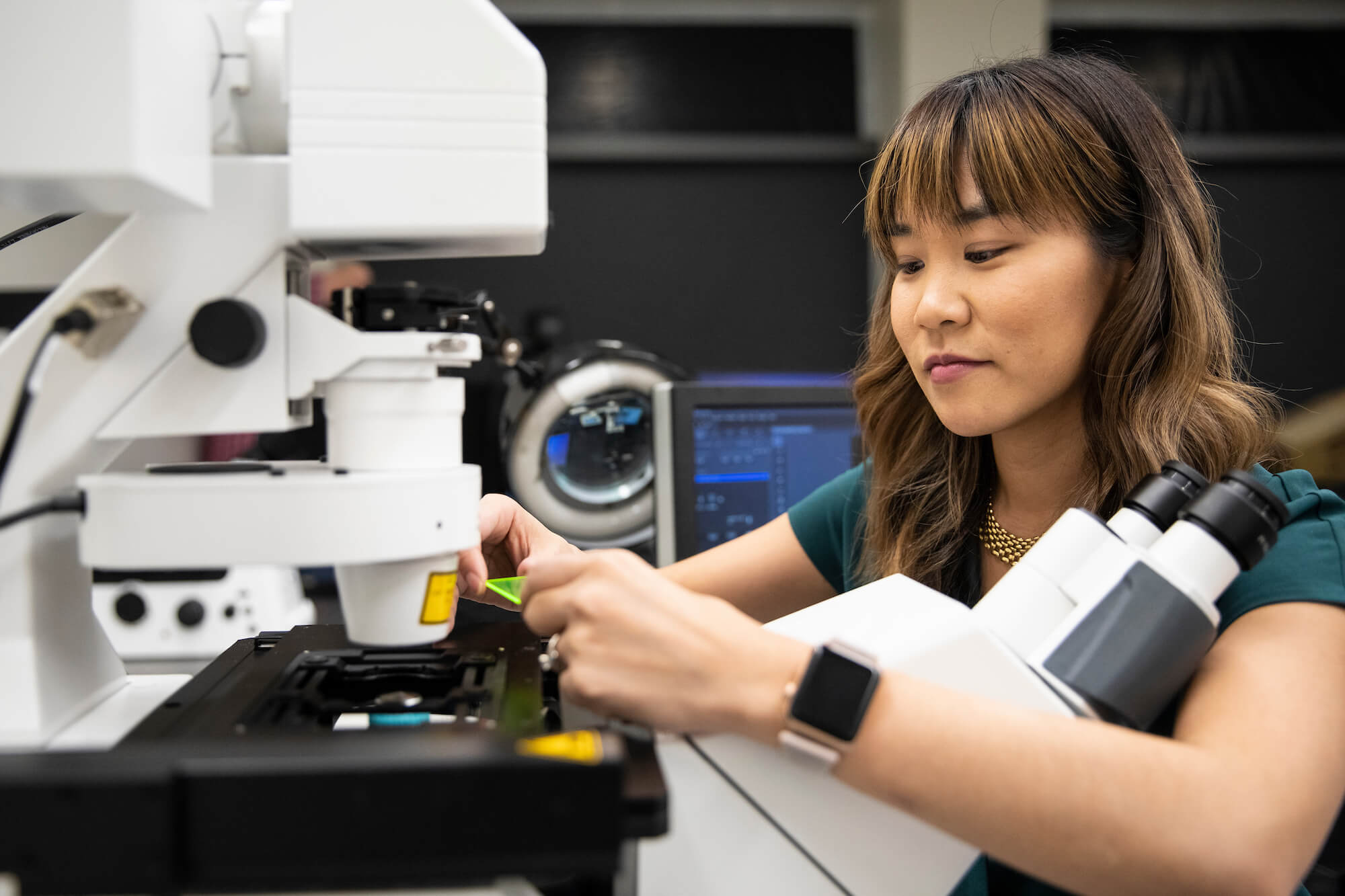
One leading scientist in this field, Dr. Michelle Itano, is developing and supporting state-of-the-art microscopy techniques to explore how viruses like COVID-19 infect cells. She’s part of the inaugural class of CZI Imaging Scientists, a program that connects, supports, and empowers scientists to develop new tools and techniques to advance a wide range of scientific research. Recently, we had the chance to speak with Dr. Itano, director of the UNC-Chapel Hill Neuroscience Microscopy Core, about the state of the field, her collaborations with other CZI Imaging Scientists, and her hopes for the future of imaging. The following are excerpts from that conversation, lightly edited for content and clarity.
What is imaging and why is it important?
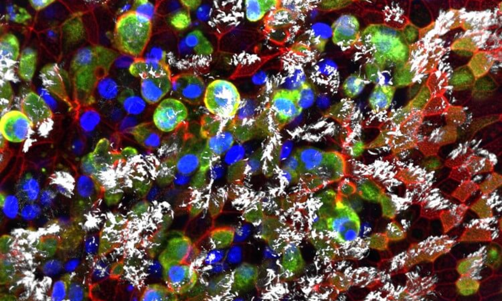
Fundamentally, the science of imaging is about seeing something that the human eye could never see alone. The type of imaging that I do is focused on microscopy, which involves using microscopes to uncover both the structure and function of any biological sample.
The thing that makes imaging so exciting is that it allows us to better understand why certain things are happening at a cellular or even molecular level. Whenever you take an image, there’s a huge amount of data there—and for a long time, it was more data in there than you could ever analyze. We help people get those images, and then pull out as much information as they can from them.
For example, right now, my core facility has been focused on supporting research on COVID-19 and continuing other basic research in spite of the impacts of COVID-19. For decades, UNC has had some of the leading labs that have worked on coronaviruses. Imaging helps us because you can see which cells get infected, and then you start asking why. How is it spreading? Why is that cell more infected than the one next to it? It’s been powerful to see imaging impact a project that really had to start from the ground up and required lots of different research collaborations.
What’s the current state of the field? Have there been any big developments in recent years?
Some people have called this time the golden period for imaging. You can’t open a big journal these days without finding multiple major papers suggesting new imaging methods to see things that people never could before, with things that are much faster or smaller than we ever thought we’d be able to measure.
There is so much creativity in the imaging world; it really is an incredibly exciting time, because barriers that we assumed were always going to be there are now being broken.
We’ve been struck by how broad the imaging community is, and the different types of scientists that it includes. How important is collaboration with different scientific disciplines for the imaging work that you do?
The way I was trained, I was taught that anybody, including undergraduates, could and should contribute to novel science. In my mind, it doesn’t matter if you’re a student, an MD, or a mathematician—you have a perspective that you can bring that can push us all to new levels of understanding.
In the graduate courses that I teach, it’s been key to have students from different disciplines. We have students from the clinical side who work with autistic children, and from the chemical side who study how membranes are formed. Students are studying everything from plants to human brain samples, and they know techniques that are not typically imaging-related—but could be adapted to make this work better and more accurate.
Imaging can use all of those perspectives, and you find that these researchers, even though their specific scientific questions may not be the same, can use each other’s expertise in a way that furthers everyone’s goals. So often, just getting people to the table and brainstorming and leaving behind the assumptions that they have from their own research project, it helps us see an old problem in new ways.
You’re part of the CZI Imaging Scientists program. What has the program been like? Have you had a chance to collaborate with any of the other 16 Imaging Scientists?
It’s been amazing. Whenever we get together, it’s always so empowering to hear what people are working on and see how open they are to collaboration. Many of the things that go into furthering our community, like job shadowing or training, have never been possible before. Having that space has been amazing.
I have several ongoing collaborations with other CZI Imaging Scientists, and those have been some of my favorite meetings during this time. It’s been great to reach out to this community and learn from what they’ve developed and help bring that back to our community at UNC.
For example, I have a lot of ongoing meetings with CZI Imaging Scientist Caterina Strambio-De-Castillia. We’re both really interested in having better tools to reproducibly pull all of the information from an image, since we need that to know how useful the image is for a certain experiment. Working with her and seeing how she is making tools for metadata has been amazing, because I can bring her insights back to my staff, and then they share their perspectives back to her. We’re able to see where people get stuck using the tools, and then work together to solve those issues.
What gives you hope about this field going forward?
I really appreciate how open the community is. The imaging community has jumped onto many open science platforms, and has started sharing specific details from their research. People are collaborating, and wanting to share data across platforms, and combine their knowledge from all these different disciplines.
Also, the fact that the field is so creative. That you will often come across something that you never imagined possible, like somebody making a totally new type of objective or software or imaging technique that pushes the field in a completely unexpected direction. That’s par for the course for imaging, and that’s what keeps me so excited about it.